Cross Section Of A Compact Bone - Tiedosto:Compact bone - ground cross section.jpg - Wikipedia - Osteocyte processes lie in tiny canals (canaliculi) in the bone matrix.
byAdmin-
0
Cross Section Of A Compact Bone - Tiedosto:Compact bone - ground cross section.jpg - Wikipedia - Osteocyte processes lie in tiny canals (canaliculi) in the bone matrix.. An estimated 10 percent of an adult's skeleton is replaced each year. The innermost layer of membrane is made up of. These are mostly compacted bone with little marrow and include most of the bones in the limbs. Between the rings of matrix, the bone cells (osteocytes) are located in spaces called lacunae. The two layers of compact bone and the interior spongy bone work together to protect the internal organs.
Haversian systems comprise concentric rings of bone around a central channel or haversian canal. Compact bone is very hard and strong. Sclerostin inhibits bone formation mostly by antagonizing lrp5/6, thus inhibiting wnt signaling. Cross section of compact bone. The two layers of compact bone and the interior spongy bone work together to protect the internal organs.
Cross-section of human femur showing trabecular and ... from www.researchgate.net Compact bone makes up the dense outer layer of bones. The remainder is spongelike cancellous bone. Compact bone is very hard and strong. Osteocyte processes lie in tiny canals (canaliculi) in the bone matrix. Magnification view of compact bone tissue. Don't assume that the cross sectional area is the same no matter where you cut. Dry bone is cut and polished before mounting on a slide. In the last decade, considerable technological improvements have been made to repair damaged bones and tissue, such as bone cross sections with implants for microscopic examinations.
Compact bone consists of closely packed osteons or haversian systems.
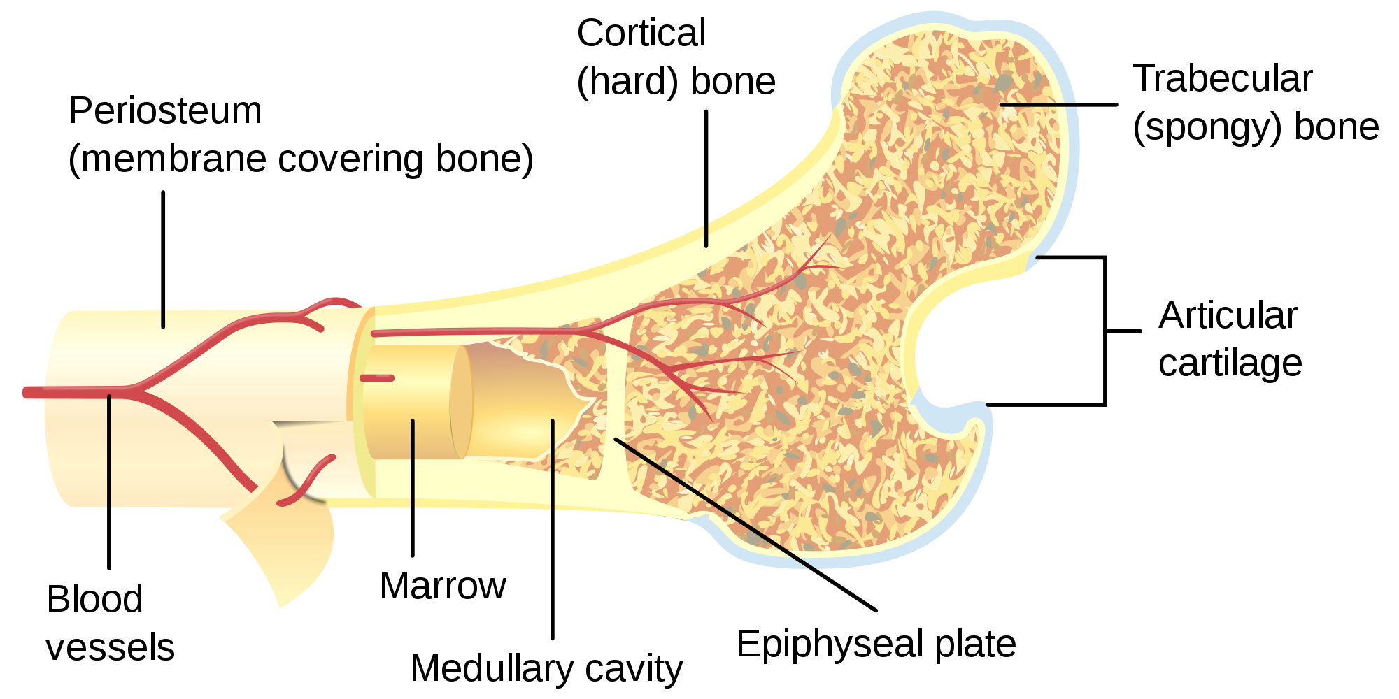
A cross section of a human long bone. The connection point for the periosteum. Select different colors for the. They build the entire picture, improve your understanding, consolidate the information and facilitate recall. To know the structures of a synovial joint and a symphysis joint (intervertebral disc). Don't assume that the cross sectional area is the same no matter where you cut. In a cross section of a bone we can see two types of bone tissue: Compact bone, makes up the dense material in a long section of a bone. Compact bone makes up the dense outer layer of bones. Cross section of compact bone. Bone decalcification is the removal of the mineral component using an acid, leaving the bone soft and easy to cut. This is a short tutorial using blender 2.8 that shows how to create a bone cross section and using images to create the textures. The two layers of compact bone and the interior spongy bone work together to protect the internal organs.
The outlined area is a cross section of an osteon of compact bone. (b) in this micrograph of the osteon, you can clearly see the concentric lamellae and central canals. This image shows compact bone in cross section. There are two ways to study bone histology. Compact bone consists of closely packed osteons or haversian systems.
Over 13 Facts About Bones-Medivizor from medivizor.com These are abundant and characteristic of compact bone. Bone decalcification is the removal of the mineral component using an acid, leaving the bone soft and easy to cut. (b) in this micrograph of the osteon, you can clearly see the concentric lamellae and central canals. Their course follows the main axis of long bone. Magnification view of compact bone tissue. There are trabeculae in spongy bone which gives its sponge like appearance. From wikimedia commons, the free media repository. Bones that are longer than they are wide are called long bones.
A cross section of a human long bone.
The osteon consists of a central canal called the osteonic (haversian) canal, which is surrounded by concentric rings (lamellae) of matrix. A central tube called a haversian canal typically runs in the same path as the length of the bone. In the center of each osteon is the central canal, a space that houses blood vessels and nerves that supply bone. Jump to navigation jump to search. Cross section of compact bone. They build the entire picture, improve your understanding, consolidate the information and facilitate recall. Their course follows the main axis of long bone. Bone decalcification is the removal of the mineral component using an acid, leaving the bone soft and easy to cut. Compact bone, also known as cortical bone, is a denser material used to create much of the hard structure of the skeleton. The outlined area is a cross section of an osteon of compact bone. To know the structures of a synovial joint and a symphysis joint (intervertebral disc). Its functional unit is the osteon. Select different colors for the.
In a cross section of a bone we can see two types of bone tissue: Select different colors for the. Canaliculi allow the passage of interstitial fluid between the central canal and the lacunae housing osteocytes. There are trabeculae in spongy bone which gives its sponge like appearance. From wikimedia commons, the free media repository.
Cross Section Of Bones / Solved: BONE TISSUE: Compact Bone ... from rlv.zcache.co.uk The outlined area is a cross section of an osteon of compact bone. This is a short tutorial using blender 2.8 that shows how to create a bone cross section and using images to create the textures. These are mostly compacted bone with little marrow and include most of the bones in the limbs. They consist of a long shaft with two bulky ends. The two layers of compact bone and the interior spongy bone work together to protect the internal organs. Compact bones make up 80 percent of the human skeleton; The spongy and compact bone tissue in the cross section of a skull bone. Between the rings of matrix, the bone cells (osteocytes) are located in spaces called lacunae.
Select different colors for the.
Concentric layers of bone cells (osteocytes) and bone matrix surround. Dry bone is cut and polished before mounting on a slide. This is a short tutorial using blender 2.8 that shows how to create a bone cross section and using images to create the textures. This is a cross section through decalcified bone. From wikimedia commons, the free media repository. Don't assume that the cross sectional area is the same no matter where you cut. In the last decade, considerable technological improvements have been made to repair damaged bones and tissue, such as bone cross sections with implants for microscopic examinations. In the center of each osteon is the central canal, a space that houses blood vessels and nerves that supply bone. The innermost layer of membrane is made up of. Their course follows the main axis of long bone. Bone decalcification is the removal of the mineral component using an acid, leaving the bone soft and easy to cut. To know the structures of a synovial joint and a symphysis joint (intervertebral disc). A diagrammatic view of a cross section of bone.
Jump to navigation jump to search cross section of a bone. (b) in this micrograph of the osteon, you can clearly see the concentric lamellae and central canals.